OAK BROOK, Unwell. – In relation to detecting numerous lung ailments from chest X-rays, human radiologists nonetheless have the sting over synthetic intelligence, a brand new examine reveals.
The examine concerned researchers pitting AI towards human radiologists to diagnose three particular lung ailments from over 2,000 X-ray scans of aged sufferers. The outcomes highlighted that AI instruments recognized a considerably increased variety of false positives — incorrectly diagnosing a illness that wasn’t really current — in comparison with their human counterparts.
This analysis reinforces the concept whereas AI can play a supportive position in medical diagnoses, it’s nonetheless early in its developmental levels.
The Danish staff from Herlev and Gentofte Hospital in Copenhagen in contrast the diagnostic abilities of 72 radiologists towards 4 totally different AI instruments utilizing 2,040 grownup X-rays taken over two years from numerous Danish hospitals in 2020. From these X-rays, roughly a 3rd (669 scans or 32.8%) introduced not less than one vital discovering.
Whereas permitted AI instruments have gotten more and more widespread as a result of a worldwide scarcity of radiologists, their real-world efficacy stays a subject of debate. The chest X-rays within the examine had been examined for 3 prevalent situations: airspace ailments (like pneumonia or lung edema), pneumothorax (collapsed lung), and pleural effusion (fluid buildup across the lungs).
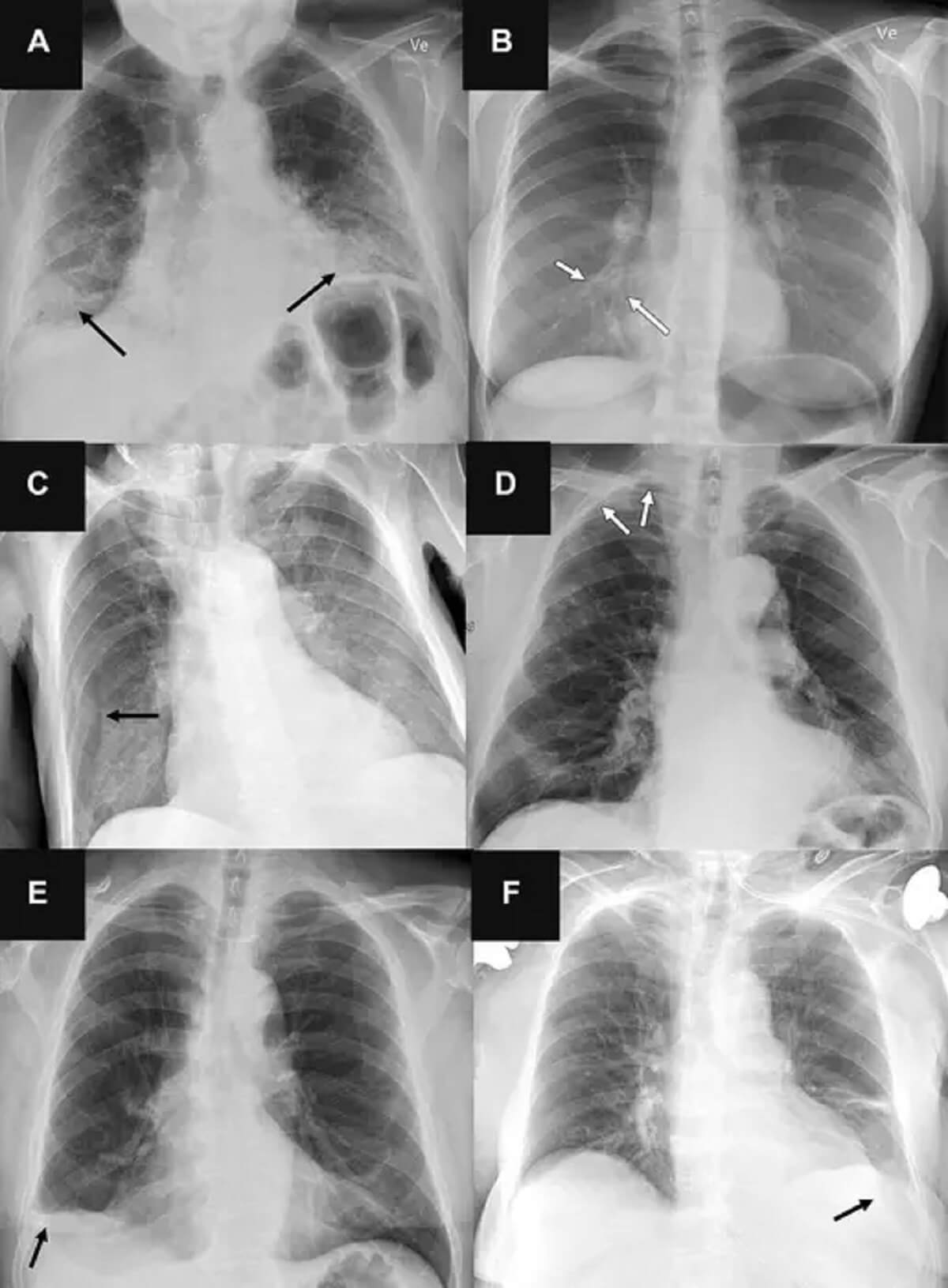
The AI instruments posted sensitivity charges between 72 and 91 % for airspace ailments, 63 to 90 % for pneumothorax, and 62 to 95 % for pleural effusion. Nonetheless, they generated extra false positives than the human radiologists, particularly when figuring out airspace ailments.
“Chest radiography is a standard diagnostic software, however vital coaching and expertise is required to interpret exams accurately,” says Dr. Louis Plesner, the examine’s lead researcher and a radiologist at Herlev and Gentofte Hospital, in a media release. “Whereas AI instruments are more and more being permitted to be used in radiological departments, there’s an unmet must additional check them in real-life scientific eventualities.”
The scientists stress that whereas AI can help in figuring out common chest X-rays, relying solely on it for diagnoses is untimely. The staff emphasizes that radiologists typically outshine AI in numerous affected person eventualities, suggesting that AI’s present state is much from prepared for particular person diagnoses. They consider that AI’s subsequent iteration, which may probably mix picture critiques with sufferers’ scientific histories and former imaging, could be far simpler, although such programs are but to be developed.
“An AI system working independently with the present charge of false positives is impractical. Whereas AI can determine common chest X-rays, it shouldn’t be solely trusted for making diagnoses,” concludes Dr. Plesner.
The examine is revealed within the journal Radiology.
South West Information Service author James Gamble contributed to this report.